Bile Duct Stones: Symptoms, Causes, and Modern Treatment Options

Need expert consultation? Book an appointment with Dr. Babu Elangovan.
Book AppointmentThe sudden onset of severe upper abdominal pain can be a frightening experience. While many people are familiar with gallbladder stones, a related and often more serious condition involves stones that find their way into the bile ducts. Known medically as choledocholithiasis, bile duct stones can obstruct the normal flow of bile, leading to debilitating pain, jaundice, and potentially life-threatening infections.
Understanding bile duct stones symptoms and treatment is essential for anyone experiencing unexplained digestive issues or those already diagnosed with gallstones. Because the biliary system is closely linked with the liver and pancreas, managing these stones requires specialized expertise.
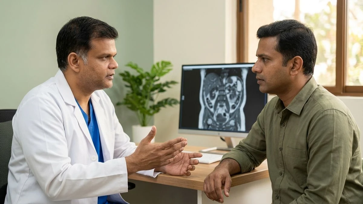
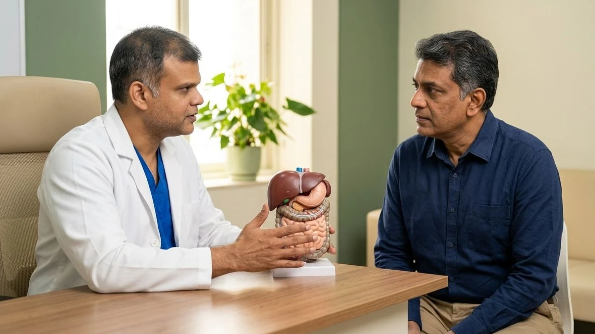
Dr. Babu Elangovan, a senior Surgical Gastroenterologist & Liver Transplant Surgeon in Chennai with over two decades of clinical experience, specializes in treating complex hepato-pancreato-biliary (HPB) disorders. Through his dedicated single-surgeon care model, patients receive consistent, personalized evaluation and treatment from their initial consultation through to complete recovery.
What Are Bile Duct Stones?
To understand how bile duct stones form and cause complications, it helps to understand the anatomy of the biliary system. The liver continuously produces bile, a digestive fluid that helps break down fats. This bile travels through a network of small tubes into the common bile duct (CBD). A portion of this bile is diverted into the gallbladder, a small pear-shaped organ that stores and concentrates it until it is needed after a meal.
When stones block these pathways, the system cannot function properly. Bile duct stones are classified into two main types based on where they originate:
- Secondary Bile Duct Stones: These are the most common type. They form initially in the gallbladder and subsequently migrate through the cystic duct into the common bile duct. They are typically composed of cholesterol.
- Primary Bile Duct Stones: These stones form directly within the bile ducts themselves. They are usually pigment stones, which develop when there is slow bile flow (stasis), structural narrowing (strictures) of the ducts, or chronic bacterial infections within the biliary system.
Regardless of their origin, stones lodged in the common bile duct restrict or completely block the flow of bile into the small intestine, leading to pressure buildup and systemic symptoms.
Gallbladder Stones vs. Bile Duct Stones: Key Differences
It is common to confuse gallbladder stones (cholelithiasis) with bile duct stones (choledocholithiasis). While they are closely related, their clinical presentation, risks, and urgency of treatment differ significantly.
| Feature | Gallbladder Stones (Cholelithiasis) | Bile Duct Stones (Choledocholithiasis) |
|---|---|---|
| Primary Location | Inside the gallbladder | Inside the common bile duct (CBD) |
| Pain Characteristics | Episodic pain (biliary colic) after heavy meals; usually subsides within a few hours | Persistent, intense pain that may not subside easily |
| Risk of Jaundice | Very low (unless a large stone compresses the duct externally) | High; obstruction directly blocks bilirubin clearance |
| Infection Risk | Cholecystitis (gallbladder inflammation) | Cholangitis (life-threatening bile duct infection) |
| Impact on Pancreas | Low risk unless a stone migrates | High risk of triggering acute gallstone pancreatitis |
| Primary Treatment | Laparoscopic gallbladder removal (cholecystectomy) | Endoscopic clearance (ERCP) or common bile duct exploration |
If you are experiencing symptoms pointing to either of these conditions, early evaluation is key to avoiding acute complications. Request a consultation with Dr. Babu Elangovan at his Chennai consulting locations for an accurate diagnosis and clear treatment pathway.
Recognizing Bile Duct Stones Symptoms
A stone resting quietly in the bile duct may not cause any symptoms initially. However, once a stone obstructs the flow of bile, symptoms develop rapidly and can become severe. Recognizing these signs early is crucial for preventing serious complications.
Biliary Colic (Severe Abdominal Pain)
The most common symptom is a sudden, sharp, or cramping pain located in the upper right side of the abdomen, just beneath the ribs. This pain can also be felt in the center of the abdomen (epigastrium) and may radiate to the right shoulder blade or back. Unlike temporary indigestion, biliary colic is usually constant, lasts for several hours, and is often accompanied by nausea and vomiting.
Obstructive Jaundice
When a stone blocks the common bile duct, bile cannot drain into the intestine. Consequently, a yellow pigment called bilirubin builds up in the bloodstream and deposits in body tissues. This causes:
- Yellowing of the skin and the whites of the eyes (sclera)
- Dark, tea-colored urine, as the kidneys attempt to filter out the excess bilirubin
- Pale, clay-colored stools, because bile pigment is no longer reaching the digestive tract to give stool its normal brown color
- Generalized skin itching (pruritus) caused by accumulated bile salts
Acute Cholangitis (Biliary Infection)
An obstructed bile duct is highly susceptible to bacterial infection. This condition, known as acute cholangitis, is a medical emergency. It is classically identified by a triad of symptoms known as Charcot’s Triad:
- Fever, often high-grade and accompanied by chills or rigors
- Right upper quadrant abdominal pain
- Jaundice
If left untreated, this infection can lead to sepsis, confusion, and low blood pressure (constituting Reynolds' Pentad), requiring immediate emergency intervention.
Gallstone Pancreatitis
The common bile duct and the pancreatic duct join together before emptying into the small intestine. A stone lodged near this shared opening can block the pancreatic duct, causing digestive enzymes to back up and inflame the pancreas. This results in severe, boring abdominal pain that radiates straight through to the back, accompanied by persistent vomiting and rapid heart rate.
Why Do Bile Duct Stones Form? (Causes & Risk Factors)
Understanding why these stones form can help patients manage their metabolic health and prevent recurrences. The primary causes and risk factors include:
- Gallbladder Disease: Having active gallstones is the single greatest risk factor for developing secondary bile duct stones.
- Biliary Stasis: Conditions that slow down or obstruct the flow of bile—such as biliary strictures (narrowing of the ducts), cysts, or scarring from previous surgeries—provide an environment where bile can crystallize into stones.
- Biliary Tract Infections: Chronic bacterial infections in the bile ducts alter the chemical composition of bile, promoting the formation of pigment stones.
- Dietary and Metabolic Factors: Rapid weight loss, prolonged fasting, obesity, and diets high in refined cholesterol can alter bile saturation, increasing stone risk.
- Age and Gender: Biliary stones are more common in individuals over the age of 40 and affect women more frequently than men, partly due to hormonal influences on bile composition.
How Are Bile Duct Stones Diagnosed?
When a patient presents with symptoms suggesting a biliary obstruction, Dr. Babu Elangovan utilizes a step-by-step diagnostic approach to locate the stone, assess the degree of blockage, and check for signs of infection or pancreatic involvement.
1. Advanced Blood Investigations
- Liver Function Tests (LFTs): These tests measure enzymes and pigments in the blood. Characteristically, obstructive stones cause a significant rise in bilirubin, Alkaline Phosphatase (ALP), and Gamma-Glutamyl Transferase (GGT).
- Complete Blood Count (CBC): An elevated white blood cell count indicates an active infection like cholangitis.
- Amylase and Lipase Levels: Elevated levels of these pancreatic enzymes point to gallbladder-induced pancreatitis.
2. Abdominal Ultrasound
An ultrasound is almost always the first imaging test performed. While it is highly reliable for detecting stones within the gallbladder, it may not always show a stone hidden deep inside the common bile duct. However, it can reliably show if the bile ducts are dilated (stretched), which is a strong indirect sign of a blockage.
3. Magnetic Resonance Cholangiopancreatography (MRCP)
MRCP is a specialized, non-invasive MRI scan that produces detailed, high-resolution images of the liver, gallbladder, bile ducts, and pancreatic duct. It is highly accurate for identifying the exact size, number, and location of stones within the ducts without requiring any radiation or invasive instruments.
4. Endoscopic Ultrasound (EUS)
In cases where stones are very small or difficult to see on standard scans, an Endoscopic Ultrasound may be recommended. This minimally invasive procedure involves passing a thin, flexible tube with a tiny ultrasound probe at its tip down the esophagus. It allows the specialist to obtain incredibly detailed images of the lower bile duct from inside the digestive tract.
5. Endoscopic Retrograde Cholangiopancreatography (ERCP)
ERCP is a specialized procedure that combines endoscopy and X-ray imaging. While it can be used for diagnosis, it is primarily reserved as a therapeutic procedure to remove stones once they have been identified by non-invasive scans like MRCP.
Modern Treatment Options for Bile Duct Stones
Once diagnosed, bile duct stones require prompt treatment to clear the blockage and prevent severe complications. The choice of treatment depends on the patient's overall health, the size and location of the stones, and whether the gallbladder is also affected.
Dr. Babu Elangovan’s extensive training in laparoscopic, robotic, and therapeutic endoscopic procedures ensures that patients have access to the most appropriate, minimally invasive treatment options.
Endoscopic Retrograde Cholangiopancreatography (ERCP)
For the vast majority of patients, ERCP is the preferred, first-line method for clearing bile duct stones.
- How it works: Under sedation, a specialized side-viewing endoscope is guided through the mouth, down the esophagus, through the stomach, and into the duodenum (the first part of the small intestine) where the bile duct opens.
- Stone Clearance: A small wire is inserted into the duct opening, and contrast dye is injected to visualize the stones under X-ray. The surgeon performs a minor incision at the muscle ring surrounding the duct opening (sphincterotomy) to widen it. Specialized baskets, balloons, or lithotripsy (stone-crushing) devices are then inserted to grasp and sweep the stones out into the intestine, where they pass naturally through the digestive tract.
- Stent Placement: If the stones are exceptionally large or if there is residual swelling, a temporary plastic or metal tube (stent) may be placed to ensure bile keeps draining freely until a definitive plan is executed.
To learn more about endoscopic biliary interventions, explore our dedicated guide on therapeutic GI endoscopy.
Surgical Common Bile Duct Exploration (LCBDE)
In some clinical scenarios, an endoscopic ERCP may not be feasible or successful. This can occur if the stones are very large or impacted, if there are anatomical variations from previous surgeries, or if the patient has a severe diverticulum near the duct opening.
In these cases, a surgical clearance is performed. Utilizing advanced laparoscopic or robotic techniques, Dr. Babu Elangovan accesses the common bile duct through tiny, keyhole incisions, opens the duct to remove the stones directly, and then closes it over a temporary drainage tube (T-tube) or stents. This approach is highly effective and avoids the need for large, open abdominal incisions.
For more information on specialized biliary surgeries, view our section on HPB surgery.
Addressing the Root Cause: Cholecystectomy
If the bile duct stones originated in the gallbladder, clearing the duct is only the first step. If the gallbladder is left intact, there is a high likelihood that more stones will migrate into the duct in the future, causing a recurrence of symptoms.
Therefore, once the bile duct is cleared and any inflammation has subsided, a laparoscopic cholecystectomy (gallbladder removal surgery) is typically scheduled. This is a highly routine, safe, and effective procedure that permanently resolves the source of the stones.
Read more about what to expect during gallbladder removal on our gallbladder & laparoscopic GI surgery page.
Managing Potential Complications Safely
Ignoring the symptoms of bile duct stones can lead to rapid clinical deterioration. Because Dr. Babu Elangovan is also a highly experienced liver transplant surgeon, he emphasizes the importance of protecting liver function from the long-term consequences of biliary neglect.
- Sepsis from Cholangitis: A bacterial infection in an obstructed duct can quickly enter the bloodstream, leading to systemic sepsis, organ failure, and shock. This requires urgent hospital admission, intensive intravenous antibiotics, and emergency duct drainage.
- Severe Pancreatitis: Pancreatic inflammation triggered by a migrating stone can lead to tissue damage (necrosis), fluid collections, and prolonged hospital stays.
- Secondary Biliary Cirrhosis: While rare, chronic, untreated, or long-standing low-grade bile duct obstruction over several years can lead to progressive liver scarring, permanent liver damage, and eventually liver failure.
Early intervention is key to preserving liver health. For patients with advanced liver disease resulting from long-term biliary complications, comprehensive evaluations are available through our liver transplant specialty service.
What to Expect During Recovery
Understanding the recovery process helps patients and their families prepare for a smooth transition back to daily life.
After an Endoscopic ERCP
Because ERCP is performed internally through the mouth, there are no external surgical incisions.
- Hospital Stay: Patients are typically observed for a few hours post-procedure or may stay overnight to ensure there are no signs of post-ERCP pancreatitis or bleeding.
- Diet: A clear liquid diet is started once the sedation wears off, gradually transitioning to soft foods and a normal diet over 24 to 48 hours.
- Activity: Most individuals can return to light, daily activities within a day or two.
After Laparoscopic or Robotic Surgery
If a surgical common bile duct exploration or gallbladder removal was performed:
- Hospital Stay: Patients usually remain in the hospital for 1 to 3 days to monitor pain, ensure adequate oral intake, and manage any surgical drains.
- Wound Care: The small keyhole incisions must be kept clean and dry. Stitches are typically absorbable or removed during a routine follow-up visit.
- Dietary Adjustments: A low-fat diet is recommended for the first few weeks following gallbladder removal. As the body adapts to digesting fats without a gallbladder, patients can gradually reintroduce a wider variety of foods.
- Activity: Heavy lifting and strenuous exercise should be avoided for 4 to 6 weeks to allow the deep abdominal tissues to heal completely, though light walking is encouraged from day one.
Choosing the Right Care Pathway in Chennai
When dealing with complex biliary conditions, having a single, highly qualified specialist manage your entire care pathway—from initial diagnosis and endoscopic stone clearance to surgical gallbladder removal and long-term follow-up—ensures seamless continuity of care.
Dr. Babu Elangovan provides comprehensive, patient-centered care for biliary and pancreatic disorders. With extensive training in both minimally invasive surgical techniques and advanced therapeutic endoscopy, he designs a treatment plan tailored specifically to your unique clinical situation.
If you or a loved one are experiencing symptoms of bile duct stones, early assessment matters. Request a consultation with Dr. Babu Elangovan at one of his convenient Chennai consulting locations:
- Mira Health Care (Adyar — Primary Consulting Clinic)
- Kauvery Hospital (Alwarpet)
- Capstone Clinics (Nungambakkam)
- Kumaran Hospital (Kilpauk)
- THANC Hospital (Kilpauk)
For outstation or international patients, remote video consultations are available to discuss diagnostic reports and obtain a specialized second opinion.
Book an appointment with Dr. Babu Elangovan today to receive expert, compassionate care for your biliary health.
References
- Williams, E. J., et al. "Guidelines on the management of common bile duct stones." Gut, 2008. https://gut.bmj.com/content/57/7/1004
- Buxbaum, J. L., et al. "ASGE guideline on the role of endoscopy in the evaluation and management of choledocholithiasis." Gastrointestinal Endoscopy, 2019. https://www.giejournal.org/article/S0016-5107(18)33162-4/fulltext
- European Association for the Study of the Liver (EASL). "EASL Clinical Practice Guidelines on the prevention, diagnosis and treatment of gallstones." Journal of Hepatology, 2016. https://www.journal-of-hepatology.eu/article/S0168-8278(16)30043-X/fulltext
- Maple, J. T., et al. "The role of endoscopy in the management of choledocholithiasis." Gastrointestinal Endoscopy, 2011. https://www.giejournal.org/article/S0016-5107(11)01568-1/fulltext
- National Institute for Health and Care Excellence (NICE). "Gallstone disease: diagnosis and management." NICE Guideline [CG188], 2014. https://www.nice.org.uk/guideline/cg188
Authored by